Babechuk Research Group Blog
News, updates, opportunities, & discussions
|
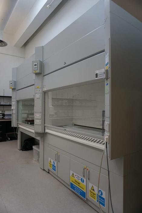
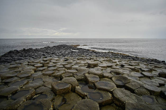
Hello folks, This blog post will briefly walk you through my time and the experiences that I had during my stay in Ireland. Mike sent me to Trinity College Dublin (TCD) as part of my BSc Honours project at Memorial, which allowed me to have a taste of what high precision trace element geochemistry is all about. I was sent there to prepare my powdered dolerite samples for inductively coupled plasma mass spectrometry (ICP-MS) analysis. Stage 0: Arrival I've finally set foot in the Emerald Isle! Everything is looking good so far. Stage 1: Introduction, Weigh-In, and Sample Digestion I met up with Cora early this day at a nice coffee shop called KC Peaches, which is right outside the complex where the clean laboratory is located. I should probably clarify that the 'clean lab' is a room inside a building called Unit 7, which in turn is inside a complex named the Trinity Enterprise Centre. Cora is the clean lab technician at TCD’s geology department. We both had a cup of coffee (this is going to be important later) and we went over the steps that I would be doing over the course of the next two weeks. We both went back inside Unit 7 to get started with work. Cora gave me a very thorough introduction of what is involved with working inside a clean lab. One might think that working in a clean lab environment simply involves wearing the standard lab coat and some gloves. Well, that is certainly not the case as working there necessitates the use of special protective equipment, which include coveralls (bunny suits), overshoes, sock covers, safety goggles, and some nitrile gloves. When one is preparing samples for ultra-trace element analysis (or when one is working in any clean lab), it is imperative to wear all these in an attempt to minimize sample contamination by skin and hair particles, as well as dust that could be brought in from outside the lab. Moreover, before entering the clean lab, it is also important to remove any metal-bearing items, such as watches and belts, as these are also potential sources of contamination. I recall Cora telling me that one of the only ways to avoid contamination in the clean lab is through telekinesis. Well, she wasn’t wrong. After Cora’s introduction to the lab, it was time for the weigh-ins. It was at this point that I wished I didn’t drink that cup of coffee earlier because both my hands were shaking like a leaf. In any case, I proceeded by weighing in approximately 100 mg of powder for every single sample (these samples were already hand milled using an agate mortar and pestle prior to traveling to Dublin) and a few standards using a spatula and some strips of weigh paper that were then used to transfer the powder into beakers made of perfluoroalkoxy alkanes (PFA) that have a high level of chemical and heat inertness. After 'weighing in' the powders, a volume of concentrated hydrofluoric acid (HF) and concentrated nitric acid (HNO3) were subsequently added to each beaker. This was, of course, done inside the fume hood for safety and cleanliness since this fume cupboard is exhausted (takes away acid vapours) and also constantly supplies clean (filtered) air. For safety reasons, it was also important to wear two sets of gloves and wrist guards when handling the HF. The samples were then sealed in the beakers and left on top of a hotplate inside the fume hood to digest for three days. I should also note that for this type of work, the concentrated acids were distilled 3 times to purify them!  Photo 3: Fancy, "hybrid" fume hoods. The one on the left is mainly used for digestions. The one on the right is mainly used for distilling acid. Note: I mentioned above that one is supposed to avoid bringing in any metal-bearing items inside the clean lab. Hint: My camera has metals in it. However, sometimes exceptions exist and Cora provided permission to take these pictures! Thanks, Cora. Stage 2: The Waiting Game Now that the samples were in the process of being digested, there is nothing else to do but relax, right? Nope. During these days, I was cleaning all sorts of lab consumables, which included stock bottles, pipette tips, test tubes, beakers, etc. Cora wasn’t joking when she told me that I would probably be spending half of my time there just cleaning consumables. In my humble opinion, maybe they shouldn’t call it a “clean lab”. Rather, it should probably be called a “cleaning lab” instead. In any case, cleaning all these consumables made me realize that good sample preparation is crucial to procuring good data. Also, when I arrived at the lab, there were already some consumables prepared for me that were ready for use. It only makes sense for me to clean as many consumables as I use. However, this is merely the bare minimum. It is always a good idea to clean more than you use. This helps keep the lab running smoothly. This will prevent instances where other people can’t finish their sample preparation due to the unavailability of properly cleaned consumables. Stage 3: Conversion During this step, the beakers were opened and put back on the hotplate to let evaporation commence. Just prior to full evaporation, concentrated HNO3 was added to each beaker and then itself evaporated. After this concentrated HNO3 was evaporated, another volume of concentrated HNO3 was added. This step converts the fluorides of the elements formed during digestion into nitrates. After the second evaporation ("dry-down") of HNO3, the samples were then dissolved in a volume of diluted HNO3 formed from adding more concentrated HNO3 and ultrapure water (Milli-Q). At this point in time, you might be wondering why HF and HNO3 are extensively used in the dissolution of silicate rocks. HF, despite being a weak acid, is one of the few chemicals that can readily dissolve silicates. This is the reason why glass cannot be used when HF is involved. However, there are a few downsides to using HF. First, you will end up losing your silicon (Si) data because HF reacts with Si to form a volatile compound called silicon tetrafluoride (SiF4) that is removed during the evaporation stages. Another downside is that extreme care must be exercised when handling HF as it is one of the most hazardous acids used in laboratories. For instance, when HF comes into contact with the human skin, it can penetrate the tissue and reach the bloodstream where it can then react with Ca and potentially lead to cardiac arrest. HF also reacts with Ca in the bones to form the insoluble calcium fluoride (CaF2). Thus, it is really crucial for anyone to wear the proper protective equipment when handling HF. HNO3 is often used in conjunction with HF because the reaction between silicates and HF forms a number of insoluble salts, especially those associated with the alkali-earth metals. A prime example of this would be CaF2. In general, the salts produced by concentrated HNO3 are quite soluble (Potts, 1992). Stage 4: Stock Solution As the heading suggests, stock solutions were made during this day. These solutions are the ones used to store the digested sample prior to diluting further for analysis. This step involved pre-filling polypropylene bottles with Milli-Q. The samples were then poured into the stock bottles and subsequently topped with more Milli-Q water to reach a sample to solution weight ratio of 1:1000 and a nitric acid solution that is approximately 2% by volume. This step sounds easy on paper, but to be honest, it was pretty taxing on the hands. To keep things clean and work with higher volumes, a squirt bottle was used, which is not necessarily the most ergonomic piece of equipment in the world. Most elements are stable in this stock solution for some time, whereas others can gradually precipitate out of solution or adsorb to the container. Stage 5: Dilution Because the ICP-MS instrument I would be using is so sensitive in detecting the elements, all samples need to be diluted much further prior to analysis! This step is relatively busy and requires a lot of care. For this step, I had to make sure that I didn’t contaminate the internal standard that Cora had concocted much earlier and for good reason too. Should the internal standard be contaminated, not only will it affect the rest of my samples, but it would also affect other people who would eventually use the same contaminated internal standard that I did. Moreover, if the pipette tip ever touched anything other than the solution it was assigned to during the dilution step, it would have to be discarded to minimize potential contamination. This step involved pipetting internal standard, followed by a small amount of stock solution, and an amount of 'carrier acid' into the same test tube. This was done for every single sample and standard that I had. The internal standard is added to every sample in the same proportion to help correct the data collected on the ICP-MS and the carrier acid helps the mixture come out to a specific dilution factor for analysis. Finally, it was time for the ICP-MS! Stage 6: ICP-MS Ah yes, I will now show you the star of the show: the lovely ThermoFisher Scientific iCap-Q mass spectrometer. This ICP-MS is a quadrupole style ICP-MS. It is a complex instrument that also looks like a mini fridge. So how exactly does the ICP-MS work? Well, it would take a long time to talk about every single detail, so I will be showing you an extremely generalized overview of what happens. The samples and standards that I prepared from Stages 1 through 5 are aspirated into the nebulizer using a peristaltic pump. The nebulizer then converts the dissolved samples into an aerosol. Argon gas flows through the ICP torch and the Ar is ionized to form a hot plasma (around 6000 degrees Celsius...that's hot!). The sample aerosol flows toward this hot argon plasma, resulting in the desolvation and atomization of the sample aerosol. The sample aerosol continues to travel through this hot plasma and it absorbs energy in the process, which leads to the release of an electron. The result is that the atoms in the sample aerosol are ionized. Well, we are halfway there. The ICP-MS is essentially an instrument that is made up of two parts – the part that makes the ions (ICP), and the part that analyzes the ions (MS). Between these two parts, there is a region called the interface. The interface is where the ion beam produced in the ICP torch is relayed to the mass spectrometer via a set of cones called the sample and skimmer cones. The ion beam passes through the sample cone and enters the interface vacuum. The ion beam then passes through the skimmer cone and then into the extraction lens, which focuses the ion beam into the quadrupole. The quadrupole filters out the ions in the beam based on their mass-to-charge ratios. The ions that are passed through the quadrupole then exit towards the detector. The detector then renders the number of ions hitting the detector into a signal that can be related to the concentration of an element in the sample. This is achieved through the use of calibration standards. As a lot of sample goes through the interface, elements are also deposited on the sample cone. This can change the diameter that the ion beam initially passes through and change the amount of ions reaching the detector. This is where our internal standard comes in! You can also think of the journey of an ion through the mass spectrometer in this way. Let’s say you invited a few friends to your house for supper after a tiring day at the gym. You and your friends (hot ion beam) reach the main door (sample cone) of your house. Not everyone can enter at the same time because of how small the main door is. Only two or three of your friends can pass through the main door and enter the foyer (interface vacuum) at any given time. Those friends of yours then proceed to pass through another door (skimmer cone), which is smaller than the main door. This time, only one person can pass through that door at any given time. Your friends ask where the kitchen is and you point toward its location. You have essentially become the extraction lens. Your friends then head over to the kitchen (quadrupole) to help you cook and prepare supper. Everyone then heads to the dining room (detector) to chow down. Everyone thanks you for the meal. You did a good job. After measuring all of my samples on the iCap-Q MS, I was able to extract all of the data. This data was in the form of a signal measured on the detector and would await to be converted to concentrations another day. The hard work was done for now. In any case, this ends the lab portion of the blog post. Stage 7: Tourism Finally, the time had come for me to become a full-fledged tourist. You can't be in Ireland without seeing some of the sights! Co. Dublin: Grand Canal Dock Samuel Beckett Bridge Guinness Brewery Trinity College Dublin Co. Wicklow Wicklow Mountains Lough Tay (Guinness Lake) Glendalough Co. Clare Cliffs of Moher Co. Antrim Giant's Causeway  Photo 21: These are the columnar basalts that we all know and love! This area is called the Grand Causeway. At the time the photo was taken, the weather was quite miserable. The photo was snapped just after a torrential downpour. I quickly realized the stuff they say about Irish weather is indeed true. At least it didn't end up like the next photo. Note: The Giant's Causeway is part of Northern Ireland, which is part of the United Kingdom. When I refer to Irish weather, I am referring to the island of Ireland as a whole, since the weather doesn't appear to recognize the border. Well, that concludes the summary of my trip to Ireland! I hope you enjoyed reading this. - Gabriel Sindol, BSc student in Earth Sciences at Memorial University of Newfoundland Acknowledgements: This research trip would not have been possible without the help of many people. I would like to thank my supervisor, Dr. Michael Babechuk, for organizing and funding the trip. I would also like to thank both Dr. Balz Kamber and Dr. David Chew from Trinity College Dublin (TCD), for being exceptionally accommodating and letting me use their laboratory facilities. Finally, I would like to thank Cora McKenna, the clean lab technician at TCD’s geology department, for teaching me all the steps and processes involved in the dissolution of silicate rocks for ICP-MS analysis and permitting me to take photos of the labs. References: Potts, P.J., 1992. A handbook of silicate rock analysis. Springer Science and Business Media, New York, 631 p.
0 Comments
Leave a Reply. |
AuthorS
Members of the Babechuk Research Group Archives
August 2019
Categories |